
Biomaterials: engineering materials that work with the human body
Every material that goes inside a human body must do something no bridge cable, aircraft fuselage, or phone screen must do: survive contact with a living system without killing it.
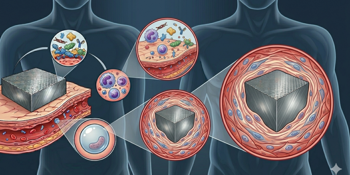
The body is not a passive environment. It’s warm, wet, chemically aggressive, and perpetually on guard. The moment a foreign material enters tissue, a cascade begins: proteins adsorb onto the surface within seconds, inflammatory cells arrive within hours, and a fibrous capsule starts forming within days. The body is trying to wall off the intruder. The engineer’s job is to either prevent that response, redirect it, or design a material that the body eventually incorporates as its own.
That’s biomaterials engineering. It’s one of the more demanding corners of the field, because the performance requirements are physiological, not just mechanical.
What makes a material biocompatible
Biocompatibility isn’t a single property. It’s a judgment about a material-host system interaction: whether a material, in a specific application, produces an appropriate host response. A material that’s perfectly acceptable in a hip joint might be toxic in a cardiac valve. Context is everything.
The first event when a material contacts blood or tissue is protein adsorption. Proteins from plasma, including fibrinogen, albumin, and immunoglobulins, arrive within seconds and coat the surface. Which proteins adsorb, in what configuration, and with what affinity, determines almost everything that follows. Cells don’t see the bare material surface: they see a layer of adsorbed proteins. The composition of that protein layer governs whether inflammatory cells are recruited, whether platelets activate, and whether tissue integration proceeds or a fibrous scar forms instead.
Surface chemistry, surface roughness, and surface energy all affect protein adsorption. The bulk properties of a material matter for structural performance, but the surface is what the body responds to. This is why surface treatments, coatings, and functionalization are central to biomaterials development: you can have a mechanically excellent material and still have a poor biological response if the surface isn’t right.
ISO 10993 is the international standard framework for biocompatibility testing. It specifies a hierarchy of tests based on the nature and duration of body contact: cytotoxicity (does it kill cells in culture?), sensitization, genotoxicity, and long-term implantation studies in animals. Clearing these tests is a prerequisite for regulatory approval. It’s a floor, not a ceiling.
Three generations of biomaterials
First generation: bioinert
The design philosophy that dominated from the 1960s through the 1980s was minimization: design a material that provokes the smallest possible biological response. Make it chemically inert. Make it mechanically strong. Hope the body tolerates it.
Titanium alloys, particularly Ti-6Al-4V, became the standard for load-bearing orthopedic implants. Titanium forms a stable oxide layer (TiO2) on its surface almost instantaneously on exposure to oxygen or water. That oxide layer is chemically inert, resists corrosion in the physiological environment, and produces a manageable inflammatory response. Ti-6Al-4V has tensile strength around 900 MPa, adequate fatigue resistance, and an elastic modulus of about 110 GPa.
That last number caused problems. Cortical bone has an elastic modulus of 10 to 30 GPa. When a much stiffer titanium stem is implanted in a femur, it carries a disproportionate share of the load. The bone, no longer being mechanically stimulated, resorbs: stress shielding. Long-term, it weakens the bone around the implant and can contribute to revision surgery.
Ultra-high molecular weight polyethylene (UHMWPE) was introduced for acetabular cup liners in hip replacements in the 1970s: low friction against metal or ceramic femoral heads, adequate wear resistance, and good biocompatibility in the bulk. The problem was wear particles. Even minimal wear generates polyethylene debris at the nanoscale. Those particles trigger a macrophage response that can lead to osteolysis (bone loss) around the implant over years. Highly crosslinked UHMWPE, introduced in the late 1990s, significantly reduced wear particle generation and extended implant lifetimes.
Second generation: bioactive
The shift toward bioactive materials came with the recognition that passive tolerance wasn’t the same as integration. A better outcome was possible if the material could actively interact with surrounding tissue.
Larry Hench at the University of Florida invented Bioglass (composition 45S5) in 1969. When implanted, Bioglass forms a hydroxycarbonate apatite layer on its surface through an ion exchange reaction with body fluids, and bone-forming cells bond directly to this layer. True bone bonding, not just mechanical interlocking. Bioglass is now used in bone graft substitutes, periodontal repair, and orthopedic coatings. Hench spent decades refining the composition and applications before his death in 2015.
Hydroxyapatite (HA) coatings on titanium implants are the most widely deployed bioactive technology. HA is the mineral phase of bone, calcium phosphate in a specific crystallographic arrangement. Plasma-sprayed HA coatings on titanium hip stems and dental implants improve osseointegration: bone grows into the coating rather than just forming a fibrous interface. The coating is mechanically weaker than the titanium substrate and can delaminate over time, which is an ongoing engineering concern.
Third generation: biodegradable and bioresorbable
If a material can be designed to degrade at a rate matching tissue regeneration, it can serve as a temporary scaffold and then disappear, leaving behind only the patient’s own tissue.
Polyglycolic acid (PGA) and polylactic acid (PLA), and their copolymers, are the most established bioresorbable polymers. PGA sutures have been used since the 1970s. Bioresorbable orthopedic screws and pins, used particularly in paediatric patients where implant removal surgery is undesirable, are made from PLA-PGA composites. The degradation rate is tunable by adjusting the PLA:PGA ratio. The clinical challenge is matching degradation rate to healing rate, and managing the local acidic byproducts of hydrolytic degradation, which can cause transient inflammatory responses.
Bioresorbable coronary stents were an ambitious application of this concept: a scaffold that holds an artery open after angioplasty, allows healing, and then disappears. Abbott’s Absorb stent was approved in Europe in 2011 and withdrawn from the market in 2017 after clinical trials showed higher rates of adverse events compared to metallic drug-eluting stents. Resorbing struts lost radial strength before the vessel remodeled. The bioresorbable stent story isn’t over, but it’s a clear example of how biological elegance and clinical outcomes don’t always align.
PEEK in spinal surgery: a case study in tradeoffs
PEEK (polyether ether ketone) is a high-performance thermoplastic used extensively in spinal surgery for intervertebral fusion cages: devices inserted between vertebral bodies after disc removal to maintain disc height and facilitate bone fusion.
PEEK’s adoption was driven by two specific advantages over the titanium cages it replaced. First, it’s radiolucent: it doesn’t block X-ray or CT imaging the way metal does, so surgeons can assess bone fusion progress through the device rather than around it. Second, its elastic modulus (approximately 3 to 4 GPa, adjustable with carbon fibre reinforcement) is closer to cortical bone than titanium’s 110 GPa, which reduces stress shielding at the endplate interface.
But PEEK is bioinert. It doesn’t bond to bone. Fibrous tissue, not bone, tends to form at the PEEK-bone interface in the absence of mechanical compression. Clinical data are mixed: fusion rates with PEEK cages are broadly comparable to titanium in most studies, but there are subgroups of patients where PEEK underperforms.
The engineering response has been to modify PEEK’s surface or composite structure. Plasma-sprayed hydroxyapatite coatings on PEEK improve osseointegration in animal models. Porous PEEK structures manufactured by 3D printing allow bone ingrowth into the material. Carbon fiber reinforced PEEK (CFR-PEEK) increases stiffness while maintaining radiolucency. The debate between PEEK and titanium for spinal fusion is active, ongoing, and unlikely to resolve cleanly, because patient anatomy, surgical technique, and implant geometry all interact with material properties in ways that are hard to control in clinical trials.
It’s a representative example of biomaterials engineering in practice: rarely a clean winner, always a set of tradeoffs, and the optimal choice often depends on the specific patient and surgeon.
Hydrogels and tissue engineering
Tissue engineering is the long game of biomaterials: grow functional tissue in the lab, transplant it into patients, and solve the organ shortage problem permanently.
The foundational paper is Langer and Vacanti’s 1993 article in Science, which articulated the strategy: seed cells onto biodegradable scaffolds, culture them in bioreactors, and implant the result. The scaffold degrades as the cells produce extracellular matrix. Thirty years later, tissue engineering has produced commercially available skin substitutes, cartilage repair products, and bladder constructs. The full-thickness complex organs that were the original vision remain largely elusive.
Hydrogels have become central to tissue engineering scaffolds. A hydrogel is a crosslinked polymer network that absorbs large amounts of water, up to 99% water content in some formulations, producing a soft, pliable material that mechanically resembles soft tissue. Natural hydrogels derived from extracellular matrix components, including alginate, gelatin, and hyaluronic acid, give cells biochemical cues similar to their native environment. Synthetic hydrogels (polyethylene glycol, polyvinyl alcohol) offer more tuneable mechanical properties and degradation rates.
Gelatin methacryloyl (GelMA) is a photocrosslinkable hydrogel widely used as a bioink in 3D bioprinting: cells are embedded in GelMA, printed into layered structures with spatial precision, then cured with UV light. The technology works well for thin, avascular tissues. Vascularization, supplying oxygen and nutrients to cells deep inside a printed construct, remains the central unsolved problem in the field.
Injectable hydrogels are a parallel development: materials liquid at room temperature that gel at body temperature or in response to other stimuli, allowing minimally invasive delivery. They’re used for drug delivery (the hydrogel degrades and releases a therapeutic over days or weeks), as void fillers in bone defects, and as carriers for cell therapy.
Where the field is going
Zwitterionic polymers, which carry both positive and negative charges in equal measure, resist protein adsorption far more effectively than PEG coatings, which have been the anti-fouling standard for years. Shaoyi Jiang’s group at Cornell developed polyzwitterionic coatings that maintain resistance to fouling for months in vivo. The mechanism is a strong hydration layer that excludes proteins sterically and electrostatically. These are entering clinical development for catheters, implants, and drug delivery particles where fouling causes device failure.
Bioelectric materials, which can conduct electrical signals and interface with neural or cardiac tissue, are a growing area. The cochlear implant is the most successful neural interface device, with over 700,000 implanted worldwide. But its electrode array is a blunt instrument. Conducting polymers (PEDOT:PSS), carbon nanotube composites, and flexible electronics fabricated on biodegradable substrates are all being developed for neural recording and stimulation with finer spatial resolution.
The endpoint most people in the field are working toward is in situ tissue engineering: implanting a scaffold that recruits the body’s own cells to regenerate tissue around it. It’s more achievable and probably more clinically robust than ex vivo tissue engineering, and it’s where the most active translational work is happening right now.
For more information or if you have any questions, please contact the author.